
Pyeloplasty
November 14, 2022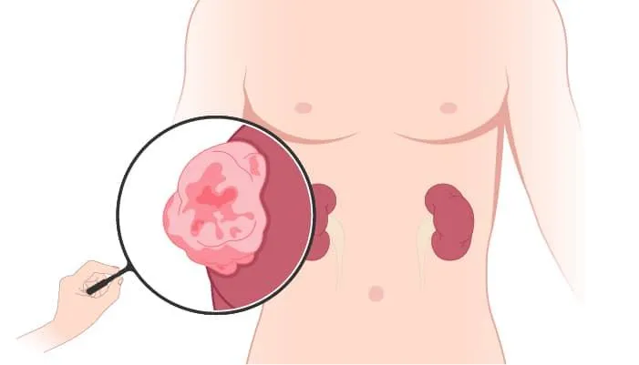
The surgical reconstruction of the renal pelvis which is a part of the kidney to drain and decompress the kidney is known as pyeloplasty. The primary objective of surgery however almost all situations would be to clear a ureteropelvic junction (UPJ) obstruction. In simple words, this procedure removes a blockage that restricts urine from reaching the bladder. In most instances, pyeloplasty primarily involves cutting out an obstructed section of the urine tube called the ureter. Because of an abnormality in how the tube developed, these blockages frequently occur right where the urine exits the kidney to travel down the tube toward the bladder. This is known as the ureteropelvic junction or UPJ.
When is Pyeloplasty done?
In children, the following conditions may require pyeloplasty:
-
Many children are born with an adynamic ureter, also known as a blocked UPJ, which has a small, tight region that is just too narrow for adequate urine to pass through. In other children, the UPJ is normal, but the blockage is caused by something outside of the ureter, such as a crossing vessel. This is referred to as an externally blocked UPJ, and it occurs more frequently in older children.
-
Rare reasons include blockages produced by polyps, tumors, or scarring.
-
When an ultrasound reveals a large kidney, many cases of clogged tubes are found before birth (hydronephrosis). Following delivery, these children are subjected to ultrasounds and other forms of imaging to discover the reason for the blockage and whether or not it will be removed.
Types of Pyeloplasty
There are various types of pyeloplasty depending on the surgical technique and incision patterns used. These include:
· Y-V Pyeloplasty
· Inverted 'U'
· Dismembered pyeloplasty techniques.
The most common type of pyeloplasty is dismembered pyeloplasty or Anderson-Hynes pyeloplasty. It is worth noting that this was mentioned about the retrocaval ureter.
What is Anderson-Hynes pyeloplasty?
Anderson-Hynes open pyeloplasty involves mobilising the upper third of the ureter and the renal pelvis, dismembering the ureter from the renal pelvis, excising the redundant renal pelvis, and reconfiguring a new PUJ. A renal vein covering the bulging pelvis can be divided, but an artery should be preserved in this situation to avoid infarction of the renal parenchyma it supplies. If such an artery exists, anastomosis is made in front of it. To splint the anastomosis, a ureteric stent is inserted. This type of procedure is now almost universally conducted using laparoscopic techniques, and in some centres, robotic assistance is used.
How it is done?
The surgery can be performed in one of three ways:
· Open surgery – It entails making a small cut, a few centimeters wide on the affected side. The skin is peeled back to allow the surgeon to see and operate directly on the child. This is usually done on newborns.
· Laparoscopic surgery - It involves making several tiny cuts (a few millimeters wide) in the abdomen. The surgeon inserts long, thin "sticks" containing tools and a camera into the tiny holes and operates from the outside of the belly.
· Robotic surgery – It is done by making several tiny cuts, around a few millimeters wide in the abdomen. To operate, the surgeon uses a computer to control the robotic arms, which move small tools beneath the skin.
Surgeons take various approaches. Neither tube is left in place in some cases. In other cases, a tube known as a "stent" may be left in place for seven to ten days to drain the ureter, or a kidney catheter tube known as a nephrostomy may be left in place for ten to twelve days.
A small drain made of soft rubber, known as a Penrose drain, may be placed beneath the incision.
Eventually, the tube or drains are removed. Although it may feel strange, removal causes only minor discomfort. A small amount of reddish-brown drainage occasionally emerges from the tubes or drains. The skin around the tubes may become red and secrete pus. This is a normal reaction to the drain and is not cause for concern.
Potential Risks
Even though this surgery is quite safe, there are risks and potential problems, as with any surgical procedure. The safety and complication rates are comparable to open surgery. Possible hazards include:
-
Bleeding: During this treatment, blood loss is often less than 100 cc, and a blood transfusion is rarely necessary.
-
Infection: To limit the potential of infection following surgery, all patients are given broad-spectrum intravenous antibiotics before the procedure. If you develop any signs or symptoms of infection following surgery, such as fever, discharge from your incision, urine frequency, discomfort, pain, or anything else that gives you concern, contact your healthcare practitioner immediately.
-
Hernias: Although hernias at incision sites are uncommon because all keyhole wounds are meticulously closed at the end of your surgery, it is possible.
-
Tissue/organ damage: Although unusual, damage to surrounding tissue and organs such as the colon, vascular structures, spleen, liver, pancreas, and gallbladder may occur.
What are the advantages of Pyeloplasty?
If pyeloplasty is required, it can protect the kidney and help it to function properly and more effectively than before it was blocked. If the treatment goes well, the youngster may be able to avoid future urinary tract infections and suffering.
If the damaged kidney is kept healthy and functioning correctly, the other kidney will not be overstressed. Having two healthy kidneys gives the child a higher chance of long-term health if the kidneys are injured or affected by another health concern.
Pyeloplasty has a high long-term success rate. However, like any surgical procedure, there is a risk of the scar tissue had returned and caused another kidney obstruction. Remember to attend all scheduled follow-up appointments. If you or the child experiences post-surgery discomforts such as nausea or vomiting, warm skin around the incision, redness around the incision, pus or drainage, severe pain that pain medications do not relieve, increased swelling around the incisions, or similar symptoms, contact surgeon or another healthcare provider as soon as possible.
No, the original ureter is approached surgically beneath the level of the obstruction, and the abnormal section is removed. The ureter is then reattached to the healthy renal pelvic tissue above.
The surgery can be performed from a variety of perspectives. In most cases, the incision will be made on your child's side. Your surgeon will discuss the best location for your child's incision with you. All surgical sutures, or stitches, will dissolve. If a tube is inserted, one skin suture is occasionally removed 10 days or so after the operation.
NOTICE BOARD
CONTACT US
FOR PARTNERSHIP
CONTACT US
FOR PARTNERSHIP
.svg)
.svg)
.svg)
.svg)


 Book Appointment
![]()