
Retinal Detachment Treatment & Diagnostics in Sadashiv Peth, Pune
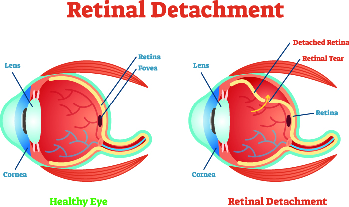
Retinal detachment is a condition in which the retina present at the back of the eye is pulled away from its normal position. It separates your retinal cells from the blood vessels that provide them with nourishment and oxygen. In simple terms, the retina is pulled away from the supportive tissue. The longer you wait to treat this, the greater is the risk of losing your vision in the affected eye permanently.
Types/Classification
There are three types of retinal detachment:
- Rhegmatogenous - Caused by a retinal tear, this is the most common type of retinal detachment. One of the main causes of this is age as the vitreous gel filling the eyeball is pulled away from the retina. It can also be caused because of surgery, nearsightedness, or an eye injury.
- Tractional - In this, the scar tissue pulls on the retina. It is caused because of damage to the blood vessels because of diabetes.
- Exudative - This type of retinal detachment occurs when there is a buildup of fluid behind the retina, but no tear. The retina is pushed away from the tissues by the fluid. Common causes of this include swelling because of age-related macular degeneration, injury, or inflammation, and leaking blood vessels.
Symptoms
In most cases, retinal detachment is painless. However, there are some warning signs that occur before the retinal detachment reaches an advanced stage. You need to look out for these symptoms:
- Photopsia (flashes of light in the eyes)
- The sudden appearance of floaters (tiny speaks drifting through the field of vision)
- Blurred vision
- Reduced peripheral vision
- A shadow over the visual field
Causes
The cause of retinal detachment depends on the type you have:
- Rhegmatogenous
a. Age
b. Eye injury
c. Nearsightedness
d. Surgery
- Tractional
a. Diabetes
- Exudative
a. Swelling caused by injury, age-related macular degeneration, or inflammation
b. Leaking blood vessels
When to see a doctor
If you experience any of the above-mentioned symptoms of retinal detachment, you should call a doctor immediately. If not treated immediately, it can lead to permanent loss of vision.
Request an appointment at Apollo Spectra Hospitals, Pune
Call 1860-500-2244 to book an appointment
Risk factors
Here are a few risk factors that increase the risk of retinal detachment:
- Being older than the age of 50
- Family history
- Myopia (extreme nearsightedness)
- Previous retinal detachment
- Previous eye surgery like cataract removal
- Previous eye injury
- Previous eye diseases like lattice degeneration (thinning of the peripheral retina), uveitis, or retinoschisis.
Preparing for a test or procedure
In order to prepare for retinal detachment treatment, here is what you can do:
- Talk to the doctor about pre-appointment restrictions in terms of eating or drinking.
- List all the medications, supplements, and vitamins you are taking.
- Have someone take you home after the procedure.
Prevention of disease
Since the main cause of retinal detachment is aging, there is no way to prevent it. However, there are ways to lower the risk of developing retinal detachment caused by an eye injury. You can wear protective eye gear or safety goggles while doing risky activities. If you start to experience any symptoms, you should visit a doctor immediately. Early intervention can prevent permanent loss of vision.
Also, you should get a comprehensive eye exam regularly. It will help the doctor detect a retinal tear or detachment early and prevent it from affecting your vision.
Treatment
The type of surgery you have to treat retinal detachment will depend on how severe the condition is. Here are a few options that you have:
- Injecting gas or air into the eye - Known as the pneumatic retinopexy, this procedure involves injecting gas or a bubble of air into your eye’s vitreous cavity. When positioned properly, the bubble will push the area containing the hole against the wall of your eye. This will stop the flow of fluid behind the retina. The doctor might also use cryopexy for repairing the retinal break. The fluid that is collected under the retina will get absorbed by itself.
- Indenting the eye’s surface - Known as the scleral buckling, this procedure involves the doctor suturing silicone material to your eye’s sclera over the affected areas. The wall of the eye is indented to relieve the force caused by vitreous tugging on your retina.
- Draining and replacing the fluid - Known as vitrectomy, in this, the doctor removes the vitreous and any tissue tugging on the retina. Then, silicone oil, gas, or air is injected for flattening the retina.
Conclusion
You must know some warning signs that occur in retinal detachment symptoms. Recognizing them and seeking medical care immediately will help save your vision.
It might take several months to get improved vision after the procedure. You might need another treatment. It is also possible that you don’t recover all of your lost vision.
It will take a few weeks for the redness to go away.
It will depend on how good your vision is in the unoperated eye.
Our Doctors

DR. VANDANA KULKARNI
MBBS, MS, DOMS...
| Experience | : | 39 Yeras Experience |
|---|---|---|
| Speciality | : | Ophthalmology... |
| Super Speciality | : | Location | : | Sadashiv Peth |
Our Top Specialities
NOTICE BOARD
CONTACT US
FOR PARTNERSHIP
CONTACT US
FOR PARTNERSHIP
.svg)
.svg)
.svg)
.svg)


 Book Appointment
![]()